Categories
Microbiology

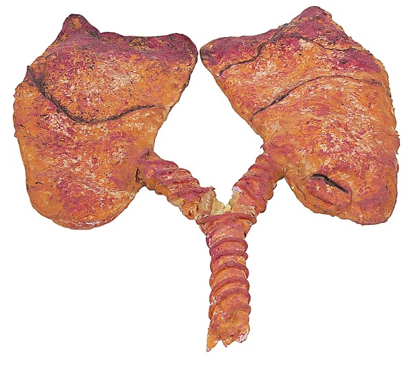
Stem cell therapy used to treat lung disease

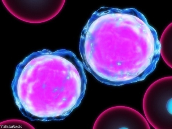
Research conducted at the Boston Children's Hospital has shown it may be possible to treat lung diseases by introducing proteins which instruct lung stem cells to produce the cell types needed to repair the injuries caused by such conditions.

A natural pathway exists in the lung which instructs stem cells to develop into specific types of cells. Researchers enhanced this natural pathway in a mouse model, successfully increasing production of alveolar epithelial cells, which line the small sacs (alveoli) where gas exchange takes place. Diseases such as emphysema and pulmonary fibrosis permanently damage these cells.
 
The researchers inhibited this pathway, causing increased production of airway epithelial cells, which are damaged by diseases that affect the lung's airways.

A 3D culture was developed to model the environment of the lung. It was found that a single lung stem cell could be made to produce alveolar and bronchiolar epithelial cells. When a protein known as thrombospondin-1 (TSP-1) was added to these cultures, they encouraged the stem cells to produce alveolar cells.

Experiments were conducted on a mouse model in which symptoms of fibrosis were induced. Endothelial cells, which line the lung's many small blood cells – and which naturally produce TSP-1 – were taken from the mice and liquid surrounding the cultured cells was injected into the mice. This process reversed the lung damage.

When the team used lung endothelial cells that lacked TSP-1 in the 3D cultures, it was discovered that the stem cells produced more airway cells. In live mice engineered to lack TSP-1, airway repair after injury was enhanced.

"When lung cells are injured, there seems to be a cross talk between the damaged cells, the lung endothelial cells and the stem cells," says Joo-Hyeon Lee, who is first author on the paper.

"We think that lung endothelial cells produce a lot of repair factors besides TSP-1," added the paper's senior author, Carla Kim. "We want to find all these molecules, which could provide additional therapeutic targets."

Categories
Microbiology

Scientists make breakthrough in developing heart tissue

Researchers have successfully engineered cardiac tissue in the laboratory that could be used as an in vitro surrogate for human myocardium and for preclinical therapeutic screenings.

Tissue which bridges the gap between animals and human subjects has been developed in the laboratory for some organs but creating such models for the heart has proved elusive. The tissue developed in the new study was generated from human embryonic stem cells and the resulting muscle is significantly similar to human heart muscle.

The research, which was published in the February 2014 issue of The FASEB Journal, was conducted at the Cardiovascular Cell and Tissue Engineering Laboratory, Cardiovascular Research Center, Icahn School of Medicine at Mt. Sinai, in New York, NY. 

Researchers cultured human engineered cardiac tissue (hECTS) for seven to ten days. They self-assembled into a long, thin heart muscle strip that pulled on the end-posts and caused them to bend with each heartbeat. The tissue was thus exercised throughout the culture process.

Spontaneous contractile activity was displayed by the hECTS, in a similar manner to the human heart, and they responded to electrical stimulation. 

Some of the functional responses known to occur naturally in the adult human heart were also induced through electrical and pharmacological interventions. Some paradoxical responses displayed by the hECTS mimicked the behavior of the immature or newborn human heart.

It was also discovered that the hECTS were able to incorporate new genetic information carried by adenovirus.

Kevin Costa, one of the researchers at the Icahn School of Medicine, commented: "This could help revolutionise cardiology research by improving the ability to efficiently discover, design, develop and deliver new therapies for the treatment of heart disease, and by providing more efficient screening tools to identify and prevent cardiac side effects, ultimately leading to safer and more effective treatments for patients suffering from heart disease."

Gerald Weissmann, editor-in-chief of the FASEB Journal, said the new tissue model could be the best yet developed on which to test therapies and model deadly diseases.

Categories
Microbiology

Molecule could protect brain from diabetes damage

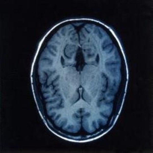
Diabetics' chances of developing dementia or Alzheimer's disease could be significantly reduced thanks to a new molecule created by researchers at the Hebrew University of Jerusalem.

High levels of sugar in the blood have been identified as a risk factor in dementia, impaired cognition, and a decline of brain function in diabetics and non-diabetics alike. The chance of diabetics developing Alzheimer's disease is twice as high as those not suffering from the disease.

A potential neuro-inflammatory pathway was discovered which could be responsible for the increased risk faced by diabetics and a possible treatment has also been identified.

A study was conducted on rats in an attempt to determine the mechanism responsible for changes in the brain due to high sugar levels. Diabetic rats were found to display high levels of enzymes called MAPK kinases, which facilitate cellular responses to a variety of stimuli, leading to inflammatory activity in brain cells and the early death of cells.

When the rats were injected with a sugar-lowering drug rosiglitazone for a month, they enjoyed a significant decrease in MAPK enzyme activity and a reduction in the inflammatory processes in the brain. The study's authors say this provides the first evidence of a functional link between high blood sugar and the activation of this specific inflammatory pathway in the brain.

A series of molecules called thioredoxin-mimetic peptides (TXM) has been developed by the study's leader, Professor Daphne Atlas, over the past few years. It mimics the action of thioredoxin and protects the cells from early death through activating inflammatory pathways. These were wound to prevent the action of MAPK kinases in animal models.

One of these molecules, TXM-CB3, significantly reduced the activity of these enzymes in the rats suffering from diabetes, reducing the accelerated brain cell death. This shows the molecule was able to traverse the blood-brain barrier and lower inflammatory processes in the animals' brains, despite the high blood sugar levels.

Professor Atlas said the discovery could lead to the development of preventive treatment in humans with high blood sugar levels.

Categories
Microbiology

Microtubule research could help develop cancer drugs

New research into the microscopic transport system that occurs within cells could help to develop new cancer treatments.

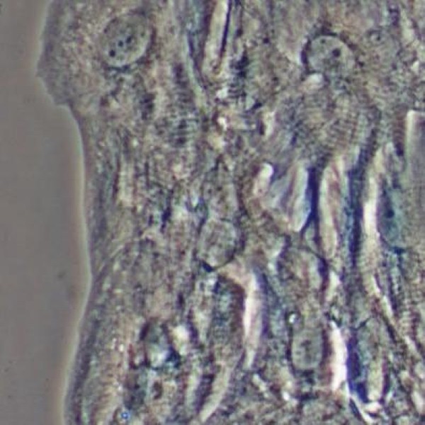
A critical point of failure in the microtubules, which act as a cellular transport system, was identified by researchers at Warwick Medical School.

Microtubules are 1,000 times thinner than a human hair and are used to transport molecules around cells. The study, published in Nature Communications, explains how these structures are targets for cancer drugs and how better treatments may be developed.

Narrow seams run down the length of microtubules, forming their weakest point. The structure dissolves if the seam cracks and splits.

Although scientists have been aware before now that microtubules have a single seam running along their length, the function of such a seam was not known. The Warwick researchers developed microtubules with extra seams in the laboratory and examined their stability using video microscopes. They found the structures become more unstable the more seams they have. 

The research has significantly altered thinking on the role of microtubules and scientists are now looking for factors inside the cell that influence the stability of microtubule seams

Some cancer therapy drugs target microtubules. Taxol, used in breast cancer therapy, prevents tumours from growing by binding to microtubules and stopping them from dissolving. This prevents microtubule tracks from remodelling themselves prior to cell division and stops them from dividing.

Professor Robert Cross, head of the research team at Warwick Medical School, explained: "It is clear that any new drugs aiming to stabilise or destabilise microtubules must target the microtubule seam. We expect this to lead us to a better understanding of the way microtubules are regulated in cells and why this sometimes goes wrong, such as in development of cancer."

He went on to say the insights the team have gained show how existing cancer drugs work and may lead to the development of more effective anti-microtubule drugs. Such drugs could have a significant impact on cancer treatment.

Categories
Microbiology

Combining therapies can help combat cancer

Scientists have discovered that two different cancer treatments developed independently can result in significantly improved outcomes when used in conjunction with one another.

Although there is a pressing demand for new cancer drugs, it is difficult for companies to develop new therapies quickly. Researchers in Ottawa tried combining existing treatments in different ways to help speed up the process.

In a report published in Nature Biotechnology, they show how two existing therapies can be used together to greater effect.

"We are very excited about this novel combination approach and are looking to move this experimental therapy into clinical trials as soon as possible," said Dr Robert Korneluk, distinguished professor at the University of Ottawa and senior scientist at the Children's Hospital of Eastern Ontario (CHEO) Research Institute. "I firmly believe that it's not a matter of 'if' this will help cancer patients – but 'when' this therapy becomes a standard of care."

Two immunotherapies are known to be effective in the fight against cancer. SMAC Mimetics targets cancer-causing genes using an IAP-based therapy. IAPs were discovered at CHEO 19 years ago. Live virus therapies, or oncolytics, is another method currently flourishing in Ottawa.

Both are currently undergoing clinical trials but neither of them, considered in isolation, has had significant effects.

However, a team of scientists led by Dr Korneluk discovered that SMAC Mimetics used in combination with a live virus (or even other non-viral immune stimulators), leads to an amplified tumour-killing effect, which overcomes the limitation of either agent working in isolation.

Dr Korneluk said the results of the experiment surpassed the researchers' expectations. Moreover, unlike conventional chemotherapy, which can produce negative side-effects, the combination of therapies did not result in any harm to the healthy tissue surrounding the tumours.

In some cases, 10,000 less virus was required to kill a cancer cell when used in combination with a SMAC Mimetic. The pioneering research could potentially save years of clinical development time and ensure patients receive treatment quicker than would otherwise occur.

Around 910 people are diagnosed with cancer every day in the UK and in 2011 more than 331,000 people in the country were diagnosed with the disease.

Categories
Microbiology

Dorsal stimulation may halt Parkinson’s symptoms

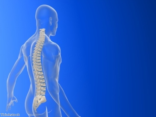
Researchers have found that long-term spinal cord stimulation may alleviate symptoms of Parkinson's disease and could protect critical neurons from injury or deterioration.

The study was conducted on rodents and builds on previous research showing that stimulation could temporarily relieve symptoms of the disease in the animals.

"Finding novel treatments that address both the symptoms and progressive nature of Parkinson's disease is a major priority," said the study's senior author Miguel Nicolelis, professor of neurobiology at Duke University School of Medicine. "We need options that are safe, affordable, effective and can last a long time. Spinal cord stimulation has the potential to do this for people with Parkinson's disease."

Parkinson's disease affects movement, muscle control and balance. It is caused by the progressive loss of neurons that produce dopamine, an essential molecule in the brain. The disease affects one person in every 500 in the UK – a total of 127,000 people.

The standard treatment for Parkinson's involves administering L-dopa, a drug that replaces dopamine but which can cause side effects and lose its effectiveness over time. Another treatment is deep brain stimulation but less than five per cent of those suffering from the disease qualify for the treatment.

In a study conducted in 2009, Nicolelis and colleagues reported they had developed a device which stimulated the dorsal column, the sensory pathway in the spinal cord carrying information from the body to the brain. When applied to the spinal column of rodents with depleted levels of dopamine, the animals' slow, stiff movements became the healthy behaviour of normal animals.

In the new study, stimulation was applied to the dorsal column twice a week for 30-minute sessions over a period of six weeks. A significant improvement in the rats' symptoms was recorded.

Stimulation was also associated with better survival of neurons and a higher density of dopaminergic innervation in two brain regions controlling movement. The loss of these causes Parkinson's in humans.

Human trials have shown dorsal stimulation may help restore motor function in humans suffering from Parkinson's, though these studies have only been conducted on a small number of people.