Categories
Microbiology

Autism linked to prenatal development

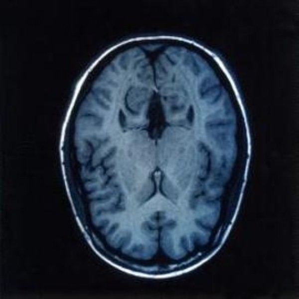
New research suggests that brain irregularities in autistic children could be traced back to prenatal development.

The study, published in the New England Journal of Medicine, shows that the origins of the architecture of the autistic brain, which contains patches of abnormal neurons, may lie in this stage of development.

"While autism is generally considered a developmental brain disorder, research has not identified a consistent or causative lesion," said Thomas Insel, director of the National Institute of Mental Health. 

"If this new report of disorganised architecture in the brains of some children with autism is replicated, we can presume this reflects a process occurring long before birth. This reinforces the importance of early identification and intervention."

Researchers from the University of California, San Diego, and the Allen Institute for Brain Science joined forces to investigate the structure of the brain's outermost layer, the cortex, in children with autism.

They analysed gene expression in postmortem brain tissue from children with and without autism, all between two and 15 years of age.

As the prenatal brain develops, neurons in the cortex differentiate into six layers, with each layer composed of different types of brain cells with specific patterns of connections.

In addition to genes associated with autism, the research team focused on genes that serve as cellular markers for each of the cortical layers.

Some 91 per cent of autistic samples lacked the markers for several layers of the cortex. For control samples, the figure was nine per cent.

Signs of disorganisation were localised in focal patches that were 5-7 mm in length and encompassed multiple cortical layers.

These signs were located in the frontal and temporal lobes of the cortex – regions that mediate social, emotional, communication and language functions. 

As disturbances in these types of behaviour are hallmarks of autism, the researchers concluded that the specific locations of the patches may be linked to the expression and severity of various symptoms in a child with the disorder.

Early treatments for children with autism may be successful because of the patchy nature of these defects – the developing brain may be able to rewire its connections by enlisting the aid of cells from neighbouring regions to bypass the pathological areas.

Categories
Microbiology

Scientists shed light on virus replication

Mathematicians and scientists from two UK universities have collaborated to shed new light on the process of viral replication during an infection.

Experimentalists from the University of Leeds and mathematicians from the University of York devised a mathematical model that gives new insights into the molecular mechanisms behind virus assembly, helping to explain the efficiency of their operation.

Researchers from the Departments of Mathematics and Biology at the University of York have developed a theoretical basis for the speed and efficiency with which viruses assemble the protective proteins for their genetic information – in this case an RNA molecule – during an infection.

The team incorporated multiple specific contacts between the genomic RNA and the proteins in the containers, along with other details of real virus infections, into a mathematical model that demonstrates how these contacts act collectively to reduce the complexity of virus formation.

They thus solved a longstanding puzzle about virus assembly – a form of Levinthal's paradox.

The process also ensures efficient and selective packaging of the viral genome and has evolved because it provides significant selective advantages to viruses that function in this way.

As a result of the research, new antiviral therapies could be developed. The team's findings could help to treat a range of diseases from HIV and Hepatitis B and C to the "winter vomiting bug" norovirus and the common cold.

Professor Reidun Twarock, a member of the York Centre for Complex Systems Analysis, said: "This truly interdisciplinary effort has provided surprising insights into a fundamental mechanism in virology. 

"Existing experimental techniques for studying viral assembly are unable to identify the cooperative roles played by all the important components, highlighting the need and power of mathematical modelling. 

"This model is a paradigm shift in the field of viral assembly."

He went on to say that the study helps elucidate the process of virus assembly and could help to develop antiviral therapies.

The findings of the research are published in the Proceedings of the National Academy of Sciences.

Categories
Microbiology

Study identifies mechanism behind kidney cancer progression

Researchers from Chicago and Beijing have identified a major mechanism behind the progression of kidney cancer and their findings could lead to new treatments for the disease. 

Their research, published in the March 20th edition of the journal Cancer Cell, shows how a shortage of oxygen, or hypoxia, created when rapidly multiplying kidney cancer cells outgrow their local blood supply, can accelerate tumor growth.

It does this by causing a nuclear cell protein known as SPOP (speckle-type POZ protein) – which normally suppresses tumour growth – to move into the cytoplasm, where it has the opposite effect.

In the cytoplasm, SPOP closes down the protective pathways that should restrict tumour growth. The team notes that the cytoplasmic overaccumulation of SPOP "is sufficient to convey tumourigenic properties onto otherwise non-tumourigenic cells."

"It becomes a vicious cycle," said the study's senior author Kevin White, professor of human genetics, and ecology and evolution, and director of the Institute for Genomics and Systems Biology at the University of Chicago.

"In people with clear-cell renal cell carcinoma, hypoxia-inducible factors enter the nucleus and target the SPOP gene. 

"SPOP gets overexpressed and misdirected to the cytoplasm, where it interferes with multiple systems designed to suppress tumor growth. This encourages tumor growth, leading to more hypoxia."

A role for SPOP as a biomarker for kidney cancer was identified by Professor White and colleagues in 2009.

They found that 99 per cent of clear cell renal cell cancers (ccRCC, the most common type of kidney cancer) had elevated SPOP levels.

The researchers hope that understanding how misplaced but not mutated SPOP contributes to cancer growth could help them identify new ways to intervene.

Work conducted on fruit flies showed that SPOP acts as a regulatory hub, influencing several cancer-related pathways.

In humans, SPOP has a profound effect, degrading multiple regulatory proteins in the cytoplasm that ordinarily serve to suppress tumour growth.

The most important was PTEN, a gene that is damaged or lost in several types of cancer. Several other tumour-suppressing proteins could be degraded by SPOP, removing additional barriers to rapid tumour growth.

SPOP may therefore turn out to be a good drug target, Professor White noted.

Categories
Microbiology

Muscle-derived stem cells used to repair nerve damage

Scientists have successfully used stem cells derived from human muscle tissue to repair nerve damage and restore function in an animal model. 

Their findings raise hopes that cell therapy of certain nerve diseases, such as multiple sclerosis, might one day be available.

Treatments for damage to peripheral nerves – those outside the brain and spinal cord – have hitherto had limited success, often leaving patients with impaired muscle control and sensation, pain and decreased function.

Researchers at the University of Pittsburgh school of medicine cultured human muscle-derived stem/progenitor cells in a growth medium suitable for nerve cells.

When prompted using specific nerve growth factors, the stem cells differentiated into neurons and glial support cells, including Schwann cells that form the myelin sheath around the axons of neurons to improve conduction of nerve impulses.

In mouse models, the researchers surgically created a quarter-inch defect in the right sciatic nerve, which controls right leg movement. They then injected the human-derived stem or progenitor cells into the defect.

After six weeks, the nerve had fully regenerated in stem-cell treated mice, while the untreated group had limited nerve regrowth and functionality.

Twelve weeks later, treated mice were able to keep their treated and untreated legs balanced at the same level while being held vertically by their tails. 

When the treated mice ran through a maze, analysis conducted on their paw prints showed an eventual restoration of gait. 

Both sets of mice experienced muscle atrophy following nerve injury but only those treated with stem cells had regained normal muscle mass by 72 weeks post-surgery.

"Even 12 weeks after the injury, the regenerated sciatic nerve looked and behaved like a normal nerve," Dr Mitra Lavasani, first author and assistant professor of orthopaedic surgery at the Pitt School of Medicine, commented. 

"This approach has great potential for not only acute nerve injury, but also conditions of chronic damage, such as diabetic neuropathy and multiple sclerosis."

The team are now trying to understand how the human-derived stem cells triggered repair of the injury and are working on delivery systems, such as gels, that could hold the cells in place at larger injury sites.

Categories
Microbiology

New method finds gene linked to heart attack risk

Researchers have identified a gene variation that has a key influence on blood lipid levels and individuals' predisposition to heart attacks.

The discovery, made by a team from the University of Michigan and the Norwegian University of Science and Technology, could help scientists to develop treatments for high cholesterol and other lipid disorders.

Significantly, the formerly unrecognised gene has been overlooked during previous attempts to find genes that affect the risk of cardiovascular disease.

While the region of DNA in which it is located had been identified as important in controlling blood lipid levels, none of them had an obvious link to these levels. An entirely new approach was needed to make the connection.

In the paper Nature Genetics, the team described how they made the discovery. They scanned genetic information in a biobank of Norwegians, focussing on variations in genes that change the way proteins function. Most of what they found turned out to be already known to affect cholesterol levels and other blood lipids.

One gene, however, was discovered to influence lipid levels. A minority of Norwegians who carried a particular change in the gene TM6SF2 were found to be healthier and at reduced risk of suffering a heart attack.

When the researchers boosted or suppressed the gene in mice, it had the same effect on the animals' blood lipid levels.

"While genetic studies that focused on common variations may explain as much as 30 per cent of the genetic component of lipid disorders, we still don't know where the rest of the genetic risk comes from," said Cristen Willer, PhD, the senior author of the paper and an assistant professor of Internal Medicine, Human Genetics and Computational Medicine & Bioinformatics at the U-M Medical School.

"This approach of focusing on protein-changing variation may help us zero in on new genes faster."

The same gene may be involved in regulating lipid levels in the liver. However, more research is needed to ascertain the exact function of the protein and whether it can be targeted with drug therapies to help treat cardiovascular disease or liver disease.

Categories
Microbiology

Study reveals microbial imbalance in Crohn’s patients

A new study has revealed a microbial imbalance in the intestinal tract of patients suffering from Crohn's disease.

The project, which involved a number of institutions, was led by investigators from Massachusetts General Hospital (MGH) and the Broad Institute. 

Their research, published in the March 12th issue of Cell Host and Microbe, reports increased levels of harmful bacteria in the gastrointestinal tract of Crohn's sufferers and reduced levels of beneficial bacteria normally found in a healthy tract.

While previous studies have linked the excessive immune response characterised by Crohn's disease to an imbalance in the normal microbial population, the exact relationship has so far not been clear.

The current study used data from the RISK Stratification Study, which was designed to investigate microbial, genetic and other factors in a group of children newly diagnosed with Crohn's disease or other inflammatory bowel diseases.

Data was also obtained from an additional group of about 800 participants in previous studies, for a total of more than 1,500 individuals.

Advanced sequencing of the microbiome in tissue samples taken from sites at the beginning and the end of the large intestine revealed a significant reduction in the diversity of the microbial population of the patients, who had yet to receive any treatment for the disease.

Compared to the control group, the Crohn's patients exhibited an abnormal increase in the levels of harmful bacteria and a drop in noninflammatory and beneficial species. There was an even greater imbalance in samples from those with more severe symptoms.

"These results identifying the association of specific bacterial groups with Crohn's disease provide opportunities to mine the Crohn's-disease-associated microbiome to develop diagnostics and therapeutic leads," said senior author Ramnik Xavier, chief of the MGH Gastrointestinal Unit and director of the MGH Center for the Study of Inflammatory Bowel Disease.

Before Crohn's is diagnosed, antibiotics are often prescribed in an attempt to alleviate symptoms. However, samples obtained from those who were taking antibiotics revealed a more pronounced microbial imbalance, suggesting this treatment could exacerbate symptoms.

Further research will focus on the function of the microbes and their products, and the interaction of these with the patient's immune system.

Categories
Microbiology

Evidence found for Turing’s morphology theory

Scientists at Brandeis University and the University of Pittsburgh have obtained evidence which supports Alan Turing's theories of morphogenesis.

While Mr Turing's accomplishments in the field of computer science are well-known, less is known about his influence on biology and chemistry.

In his only paper on biology, he put forward a theory of morphogenesis, explaining how identical copies of a single cell differentiate into an organism with separate structures.

Researchers have provided experimental evidence for Mr Turing's theory for the first time, publishing their findings in the Proceedings of the National Academy of Sciences.

The famous mathematician, who worked at Britain's code-breaking centre at Bletchley Park during the second world war, was the first to offer a theory of morphogenesis through chemistry.

He theorised that identical biological cells differentiate, change shape and create patterns through a process called intercellular reaction-diffusion.

According to the theory, chemicals react with each other and diffuse across space; they require an inhibitory agent, to suppress the reaction, and an excitatory agent, to activate the reaction. Chemically different cells are produced as a result of the chemical reaction, diffused across an embryo.

In order to test the hypothesis, scientists at Brandeis – Seth Fraden, professor of physics, and Irv Epstein, the Henry F Fischbach professor of chemistry – created rings of synthetic, cell-like structures with activating and inhibiting chemical reactions.

Mr Turing predicted six different patterns that could arise from this model and the scientists observed all of these, plus a seventh.

The researchers found that, in line with Mr Turing's theory, once-identical structures – now chemically different – also began to change in size due to osmosis.

It is hoped the research will aid the study of biological development and how similar patterns form in nature. It could also have an impact on materials science – Turing's model could help grow soft robots with certain patterns and shapes.

Last year, the Queen issued a posthumous pardon for Mr Turing, who was prosecuted for homosexuality in 1952 and committed suicide two years later.

Categories
Microbiology

Research uses familial stem cells to study AD

A team of researchers has used stem cells to help understand the processes underlying the development of Alzheimer's Disease (AD) and to investigate new treatment possibilities.

Scientists at Brigham and Women's Hospital (BWH) used stem cells derived from related family members with a genetic predisposition to the disease.

Dr Tracy Young-Pearse, corresponding author of the study recently published in Human Molecular Genetics and an investigator in the Center for Neurologic Diseases at BWH, pointed out that postmortem tissue had typically been used in previous studies of the disease.

"In this study, we were able to generate stem cells from skin biopsies of living family members who carry a mutation associated with early-onset AD. We guided these stem cells to become brain cells, where we could then investigate mechanisms of the disease process and test the effects of newer antibody treatments for AD."

Skin biopsies were obtained from a 57-year-old father with AD and his 33 year-old daughter, who is currently asymptomatic for AD. Both individuals harbour the "London" familial AD Amyloid Precursor Protein (APP) mutation, V7171.

The biopsies were submitted to the Harvard Stem Cell Institute, where the cells were converted into pluripotent stem cells. The team then directed these into neurons specifically related to a particular region of the brain which is responsible for memory and cognitive function.

The scientists showed that the APPV7171 mutation alters APP subcellular location, amyloid-beta protein generation, and then alters Tau protein expression and phosphorylation. This impacts the Tau protein's function and activity.

Amyloid-beta antibodies were then tested on the affected neurons. The team demonstrated that the secondary increase in Tau can be rescued by treatment with the amyloid-beta protein antibodies, providing direct evidence linking disease-relevant changes in amyloid-beta to aberrant Tau metabolism in living cells obtained directly from an AD patient.

 Dr Young-Pearse said the research could be useful in testing and comparing amyloid-beta antibodies, which are proving to be a promising therapeutic technique if delivered early in the disease process.

Categories
Microbiology

Researchers identify protein that helps cancer cells survive

New research conducted at the Translational Genomics Research Unit (TGen) has identified a key protein that helps prevent the destruction of lung cancer tumours, which could make radiation treatment and chemotherapy more effective.

Non small cell lung cancers (NSCLCs) account for 87 out of 100 lung cancers in the UK. Lung cancer is the second most common cancer diagnosed in the UK and the most common cause of cancer death in the UK, accounting for more than one in five deaths from the disease.

Platinum-derived chemotherapeutics, such as cisplatin, or radiation therapy are often used to treat the disease in the absence of more effective targeted therapies.

Studies previously conducted at TGen have shown that excessive activation of a cellular signaling mechanism known as TWEAK-Fn14 is linked to the survival and spread of cancer cells.

In a new study, published in the scientific journal Molecular Cancer Research, researchers found that a protein called Mcl-1 helps enable TWEAK-Fn14, which in turn helps protect NSCLC tumours from being destroyed by radiation and drugs.

"Our study demonstrates that the expression of Mcl-1 is necessary to promote the TWEAK-mediated survival of NSCLC tumor cells," said Dr Timothy Whitsett, an assistant professor in TGen's Cancer and Cell Biology Division, and the study's lead author. "By deactivating Mcl-1, we believe we can give these lung cancer patients a better response to standard therapy."

A drug called EU-5148 was used to block Mcl-1 function and halt the TWEAK-Fn14 cellular signaling mechanism. Inhibition of Mcl-1 enhanced chemo- and radio-sensitivity in NSCLC cells.

Dr Nhan Tran, an associate professor in TGen's Cancer and Cell Biology Division, and the study's senior author, said the TWEAK-Fn14 cellular pathway and the Mcl-1 protein are potential therapeutic interventions.

He added that bypassing these mechanisms would make it more difficult for lung cancer cells to evade therapies.

According to the team, more research is needed into the Mcl-1 and TWEAK-Fn14 mechanism. Clinical trials would follow, hopefully leading to more effective treatments that could reduce lung cancer mortality.

Categories
Microbiology

New technique developed to image biomolecule dynamics

Researchers at Columbia University have come closer to realising a longstanding goal of the scientific community by developing a new method of visualising small biomolecules inside living biological systems with minimum disturbance.

A general method has been developed to visualise a range of molecules such as small molecular drugs and nucleic acids, amino acids and lipids, determining where they are localised and how they function inside cells.

Fluorophores – molecules that glow when illuminated – have been used to label molecules of interest when studying their activity within cells. A fluorescence microscope is used to track and locate these molecules with high precision. This process became more common following the invention of green fluorescent protein in 1994.

Fluorophore tagging is not without its problems, however. Difficulties arise when tagging small biomolecules as the fluorophores are almost always larger or comparable in size to the small molecules of interest. As a result, they often disturb such molecules' functioning.

Assistant professor of Chemistry Wei Min's research team used an emerging laser-based technique called stimulated Raman scattering (SRS) microscopy. They combined this with a small but highly vibrant alkyne tag (C=C, carbon-carbon triple bond), a chemical bond that, when it stretches, produces a strong Raman scattering signal at a unique "frequency" (different from natural molecules inside cells). 

Using the tiny alkyne tag to label molecules avoids the problems associated with fluorophore tagging while obtaining high-detection specificity and sensitivity by SRS imaging.

The laser colours are tuned to the alkyne frequency and the focused laser beam quickly scanned across the sample, point-by-point, enabling SRS microscopy to pick up the unique stretching motion of the C=C bond carried by the small molecules. A three-dimensional map of the molecules inside living cells and animals is thus obtained.

Using this method, Min's team demonstrated tracking alkyne-bearing drugs in mouse tissues and visualising de novo synthesis of DNA, RNA, proteins, phospholipids and triglycerides through metabolic incorporation of alkyne-tagged small precursors in living cells.

The team intends to use this technique in further experiments; they are also creating other alkyne-labeled biologically active molecules for more versatile imaging applications.